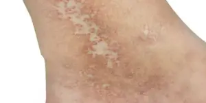
What is Atrophie Blanche?
Atrophie blanche is a dermatologic finding characterized by white-or-light- colored patches of skin in regions with darker discoloration. This is a result of necrosis and the replacement of necrotic tissue with fibrin deposits and collagen. As discussed in a previous blog, this condition would be recognized as C4b on the CEAP classification.
View our classification of venous disease blog here.
From a diagnostic coding standpoint, it is “varicose vein with ulceration.” Insurers do not use the CEAP classification system at this time, although there may be some expectation of a minimal amount of disease. From the standpoint of coding as well as the standpoint of proper management, an ulcer is an ulcer. It doesn’t matter if they had it before, have it now, or they didn’t even know they had it(as in the case of atrophie blanche). The treatment is the same; we find the perforator vein and the other points of reflux and we ablate them.

When circling the CEAP on the history or physical exam, the patient is C4, but for diagnostic coding, it doesn’t matter if the ulcer is active, inactive, or not know to the patient-it is on the spectrum of venous insufficiency with ulceration. It is dead tissue that has been replaced by scar tissue.
At Allure Medical…
We treat not only varicose veins, but we also treat “ugly legs”. These ugly legs may range from minor skin changes to atrophie blanche, and “ugly leg” patients may have active or healed known ulcerations. These patients typically will have more diseases and more likely to be overweight.
Think you might have Atrophie Blanche, or leg ulcers due to venous disease? Schedule a free leg exam now.
Already having issues? Schedule your free leg exam today!


